
Everything About The Triangle of Koch (Human Cardiac Anatomy) Triangle of koch, Cardiac
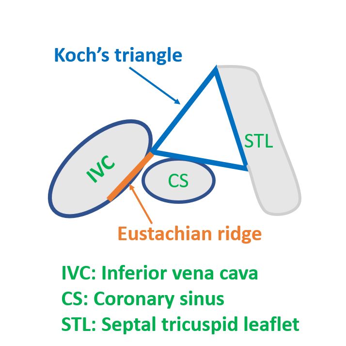
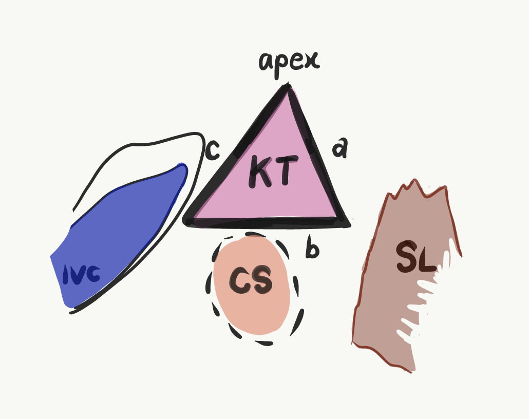
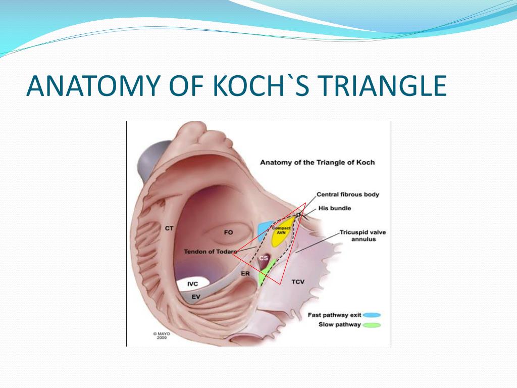
The triangle of Koch is defined by the following structures within the right atrium: (1) The ostium of the coronary sinus, posteriorly; (2) the anterior portion of the tricuspid valve annulus; and (3) the tendon of Todaro (a tendinous structure connecting the valve of the inferior vena cava ostium to the central fibrous body), posteriorly.

Pin em Triangles in anatomy
ANATOMY - the Triangle of Koch is comprised of superficial structures located in the low right atrium. Defined by three anatomical borders: Septal leaflet of the tricuspid annulus Tendon of Todaro, the apex of the Triangle, anterior structure ( His bundle catheter placement)

koch triangle heart Cerca con Google Anatomy Pinterest Heart, Simple and Pathways
Morphometry of the triangle of Koch and position of the coronary sinus opening in cadaveric fetal hearts - PMC Journal List Indian Heart J v.69 (1); Jan-Feb 2017 PMC5319009 As a library, NLM provides access to scientific literature.

Triangle of Koch. (a) Cadaveric image shows the triangle of Koch (green... Download Scientific
The mean value of the Koch's triangle area was 151.5 ± 55.8 mm 2. The 95th percentile of triangle's height (the distance from the apex to the coronary sinus) was 21.8 mm. Conclusion Mean values and proportions of triangle's sides and angles were presented. Koch's triangle showed considerable individual variations in size.

Everything About The Triangle of Koch (Human Cardiac Anatomy) Medrenaline
The coronary sinus originates from the confluence of the oblique vein (of Marshall) of left atrium and the great cardiac vein, and receives the small and middle cardiac veins, and the posterior vein of the left ventricle as tributaries. This article will discuss the anatomy and function of the coronary sinus. Contents Anatomy and course

Variable Arrangement of the Atrioventricular Conduction Axis Within the Triangle of Koch
The triangle of Koch is the target of ablation for atrioventricular node reentrant tachycardia, septal and paraseptal accessory pathways, and atypical forms of atrial flutter (32,33). The size of the triangle of Koch varies in different individuals, with a mean height of 26 mm ± 8 .
Triangle of Koch All About Cardiovascular System and Disorders
The triangle of Koch was first described and named after the German pathologist Walter Koch. Koch's triangle constitutes a target area for ablation procedures of atrioventricular nodal reentrant tachycardia or atrial flutter . innervation of the heart. atrioventricular septum.
Everything About The Triangle of Koch (Human Cardiac Anatomy) Medrenaline
Discussion on triangle of Koch and its importance while ablating supraventricular tachycardia.Triangle of Koch is situated on the right atrial aspect of the.

Everything About The Triangle of Koch (Human Cardiac Anatomy) Medrenaline
The Triangle of Koch is one of the important anatomical areas, it is located in the right atrium of the human heart, to be specific in the superficial paraseptal endocardium of the right atrium.

Schematic view of Koch triangle, prior (A) and following (B) ablation... Download Scientific
Triangle of Koch. Triangle of Koch with the compact atrioventricular (AV) node and its inferior and superior extensions. Contributed by Spyridon Koulouris, MD. Modified from Shereen. A comprehensive review of the anatomical variations in the right atrium and their clinical significance.

Triangle of Koch, Inferoseptal Recess and Inferior Pyramidal Space Download Scientific Diagram
This anatomic region is also commonly referred to as the triangle of Koch. The blood supply to the AV node is from the AV nodal branch of the right coronary artery (90%) or the left circumflex artery (10%) depending on the right or left dominant blood supply to the heart. The first septal perforator of the left anterior descending artery also.

Relationship to the conduction system
Triangle of Koch is situated on the right atrial aspect of the inter-atrial septum. It is bounded by the septal tricuspid leaflet, tendon of Todaro and the coronary sinus. The atrioventricular node is located at the apex of this triangle [1]. The initial description of the triangle was by Walter Koch in 1909 [2].

Highresolution mapping of the triangle of Koch Spatial heterogeneity of fast pathway
Introduction: There is growing use of the Todaro tendon and triangle of Koch as anatomic icons for invasive cardiac electrophysiologists. Reasons exist to doubt this validity. Methods and results: Histologic sections were prepared from 96 anatomically normal human hearts. The study area extended from the crista supraventricularis to the eustachian valve and included the AV node and His bundle.
PPT Narrow complex tachycardia PowerPoint Presentation, free download ID5020229
Koch's triangle, named after the German pathologist Walter Koch, [1] is an anatomical area located in the superficial paraseptal endocardium of the right atrium, which its boundaries are the coronary sinus orifice, tendon of Todaro, and septal leaflet of the right atrioventricular valve. [2]

Anatomic drawing of the triangle of Koch including anterior (A), medial... Download Scientific
This anatomic region is also commonly referred to as the triangle of Koch. The sinoatrial nodal artery supplies blood to the sinoatrial node, it branches off the right coronary artery in 60% of cases, whereas in 40% of cases, it comes from the left circumflex coronary artery. The blood supply to the AV node is from the AV nodal branch of the.

Pin by Kimberly Volkman on lovecho Cardiac anatomy, Diagnostic medical sonography, Anatomy
Koch's triangle, named after the German pathologist Walter Koch, is an anatomical area located in the superficial paraseptal endocardium of the right atrium, which its boundaries are the coronary sinus orifice, tendon of Todaro, and septal leaflet of the right atrioventricular valve.
